
Incubator Accessories

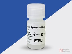
Diagnostic Biosystems BROAD SPECTRUM Broad Spectrum Negative Control
Negative controls are used to determine the specificity of an antibody. No staining should be observed when the primary antibody is omitted. This product is specifically designed to work with rabbit, mouse and goat antibodies. The Broad Spectrum Negative Control has been titered to work with Polymer-based Secondary Systems in manual and automated IHC platforms. This product is specifically designed to work with rabbit, mouse and goat antibodies. The Broad Spectrum Negative Control has been titered to work with Polymer-based Secondary Systems in manual and automated IHC platforms.

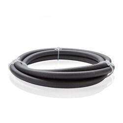
Foxx Life Sciences EZBiopure Chemically Resistant Fluoroelastomer (FKM) Supercritical Fluid Transfer Tubing 5mm (13/64") ID X 10mm (25/64") OD, 50FT

EZBiopure Fluoroelastomer (FKM) Tubing (popularly known as Viton tubing) is engineered for supercritical fluid transfer and the safe handling of highly corrosive chemicals and solvents in pharmaceutical applications. It is produced in a controlled cleanroom environment to meet stringent food and pharma-grade standards.
Features
Made and packaged in an ISO Class 7 certified cleanroom.
Chemical Resistance: Excellent compatibility with alcohols, acids, and halogenated solvents (e.g., IPA, MDC).
High Safety: Superior flame and fire resistance.
Wide Temperature Range: Operational from –15°C to +250°C.
Applications:
Corrosive chemical transfer
Chlorinated, fluorinated chemical transfer
Chemical and analytical analysis
Fuel and oil industries
Certifications
FDA 21 CFR 177.2600 Certificate
USP Class VI Certificate
Phthalates / Bis Phenol / Plasticizer free Certification
TSE/BSE Free Certification
5 years Shelf Life

Enterprise Technology Solutions Extra Capacity Reinforced Hang
Extra Capacity Reinforced Hanging File Folders with Box Bottom 2 Capacity Legal Size 1/5-Cut Tabs Red 25/Box
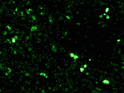
Diagnostic Biosystems C1Q-FITC C1q Complement-FITC
This antibody reacts with the human C1q complement. Traces of contaminating antibodies have been removed by solid phase absorption with human plasma proteins. This antibody is well suited for tissue section staining but may also be used for other applications.


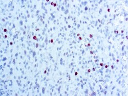
Diagnostic Biosystems PHOSPHOHISTONE H3 PHH3 Phosphohistone H3 (PHH3)
Phosphohistone H3 (PHH3) is a core histone protein, which together with other histones, forms the major protein constituents of the chromatin in eukaryotic cells. In mammalian cells, phosphohistone H3 is negligible during interphase but reaches a maximum for chromatin condensation during mitosis.Immunohistochemical studies showed anti-PHH3 specifically detected the core protein histone H3 only when phosphorylated at serine 10 or serine 28. Studies have also revealed no phosphorylation on the histone H3 during apoptosis.PHH3 can serve as a mitotic marker to separate mitotic figures from apoptotic bodies and karyorrhectic debris, which may be a very useful tool in diagnosis of tumor grades, especially in CNS, skin, gyn., soft tissue, and GIST.
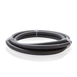
Foxx Life Sciences EZBiopure Chemically Resistant Fluoroelastomer (FKM) Supercritical Fluid Transfer Tubing 6mm (15/64") ID X 8mm (5/16") OD, 25FT
EZBiopure Fluoroelastomer (FKM) Tubing (popularly known as Viton tubing) is engineered for supercritical fluid transfer and the safe handling of highly corrosive chemicals and solvents in pharmaceutical applications. It is produced in a controlled cleanroom environment to meet stringent food and pharma-grade standards.
Features
Made and packaged in an ISO Class 7 certified cleanroom.
Chemical Resistance: Excellent compatibility with alcohols, acids, and halogenated solvents (e.g., IPA, MDC).
High Safety: Superior flame and fire resistance.
Wide Temperature Range: Operational from –15°C to +250°C.
Applications:
Corrosive chemical transfer
Chlorinated, fluorinated chemical transfer
Chemical and analytical analysis
Fuel and oil industries
Certifications
FDA 21 CFR 177.2600 Certificate
USP Class VI Certificate
Phthalates / Bis Phenol / Plasticizer free Certification
TSE/BSE Free Certification
5 years Shelf Life
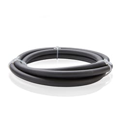
Foxx Life Sciences EZBiopure Chemically Resistant Fluoroelastomer (FKM) Supercritical Fluid Transfer Tubing 6.35mm (1/4") ID X 12.7mm (1/2") OD, 25FT
EZBiopure Fluoroelastomer (FKM) Tubing (popularly known as Viton tubing) is engineered for supercritical fluid transfer and the safe handling of highly corrosive chemicals and solvents in pharmaceutical applications. It is produced in a controlled cleanroom environment to meet stringent food and pharma-grade standards.
Features
Made and packaged in an ISO Class 7 certified cleanroom.
Chemical Resistance: Excellent compatibility with alcohols, acids, and halogenated solvents (e.g., IPA, MDC).
High Safety: Superior flame and fire resistance.
Wide Temperature Range: Operational from –15°C to +250°C.
Applications:
Corrosive chemical transfer
Chlorinated, fluorinated chemical transfer
Chemical and analytical analysis
Fuel and oil industries
Certifications
FDA 21 CFR 177.2600 Certificate
USP Class VI Certificate
Phthalates / Bis Phenol / Plasticizer free Certification
TSE/BSE Free Certification
5 years Shelf Life
Bellco Glass, Inc. EZFlow Membrane Disc Filter, 0.22µm Nylon, 90mm, 25/PK
Disc Filter with Nylon MembraneEZFlow 13mm, 25mm, 47mm, and 90mm membrane disc filters are used to retain particles or microorganisms by surface capture. The membrane disc filters are available in a nylon material or hydrophobic PTFE material with polypropylene support, offered in 0.2µm or 0.45µm pore sizes. The Nylon membrane has great chemical resistance and makes them ideal for filtering more aggressive solutions such as alcohols and DMSO. The Hydrophobic PTFE membrane is bonded to a polypropylene support and due to its extreme chemical compatibility, they are ideal for the filtration of solvents and acids or other aggressive chemicals. All materials comply with USP Class VI Standards and FDA compliances.USP Class VIFDA ApprovedSpecificationsFilter Diameter: 90mmFilter Pore Size: .2µmFilter Material: Nylon
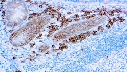
Diagnostic Biosystems CD138 CD138 (B-A38)
CD138, also known as Syndecan-1, is a member of the transmembrane heparan sulfate proteoglycan family, acts as an extracellular matrix receptor and is involved in many cellular functions, including cell-cell adhesion and cell-matrix adhesion. CD138 expression is found in both hematopoietic and non-hematopoietic cells. In the hematopoietic system, CD138 labels plasma cells. It is an excellent marker for plasmacytic differentiation within the spectrum of hematologic malignancy. Among non-hematolymphoid cells, CD138 reactivity is observed in many types of epithelial cells and stoma cells in both normal and tumor tissues.
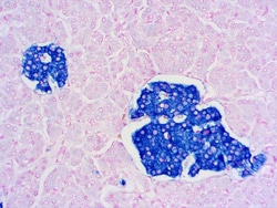
Diagnostic Biosystems SYNAPTOPHYSIN Synaptophysin (SP11)
This antibody recognizes a protein of 38kDa, identified as synaptophysin. It labels normal neuroendocrine cells of human adrenal medulla, carotid body, skin, pituitary gland, thyroid, lung, pancreas, gastrointestinal mucosa, Paneth’s cells in the gastrointestinal tract and gastric parietal cells. Neurons in the brain, spinal cord, and retina are also labeled. In combination with anti-chromogranin A and anti-NSE, anti-synaptophysin is very useful in the identification of normal neuroendocrine cells and neuroendocrine neoplasms.

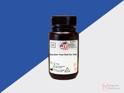
Diagnostic Biosystems NUCLEAR FAST RED FOR IHC Nuclear Fast Red
Nuclear Fast Red is a histological staining reagent suitable for visualization of nuclei in tissue sections. Nuclear Fast Red provides a red counterstain designed for optimal performance when used in immunohistochemistry.\n\nDBS’s Nclear Fast Red is intended for use in the histologic demonstration of nuclear staining. This staining technique is used to make the critical distinction between a normal nucleus and an abnormal one. When used as a counterstain in immunohstochemical procedures, Nuclear Fast Red is compatible with most commonly used chromogens including DBS HRP-Yellow, HRP-Green, HRP-Black, and DAB. Nuclear Fast Red is particularly useful with blue chromogens, such as DBS HRP-Blue.